The CLEI Center for Keratoconus, under the direction of Dr. Peter Hersh, is actively involved in research programs to develop new technologies and treatments for keratoconus and related corneal diseases.
Our dedicated research team consists of doctors and researchers who devote their time to clinical trials and other efforts dedicated to advancing patient care in keratoconus, LASIK, and other problems of the eye and cornea.
Our work has been published in some of the most prominent journals in the field. Please click the below banners below to read the full article.
Purpose: To evaluate the safety and efficacy of corneal collagen crosslinking (CXL) for the treatment of progressive keratoconus.
Conclusions: Corneal collagen crosslinking was effective in improving the maximum keratometry value and vision in eyes with progressive keratoconus 1 year after treatment, with an excellent safety profile.
Corneal collagen crosslinking affords the keratoconic patient an important new option to decrease the progression of this ectatic corneal process.
Purpose: To evaluate the safety and efficacy of corneal collagen crosslinking (CXL) for the treatment of corneal ectasia after laser refractive surgery.
Conclusions: Corneal collagen crosslinking was effective in improving the maximum K value and vision in eyes with corneal ectasia 1 year after treatment, with an excellent safety profile. CXL is the first approved procedure to diminish the progression of this ecstatic corneal process.
Purpose: To evaluate 1-year outcomes of corneal collagen crosslinking (CXL) for the treatment of keratoconus and corneal ectasia.
Conclusions: Collagen crosslinking was effective in improving vision, the maximum K value, and the average K value. Keratoconus patients had more improvement in topographic measurements than patients with ectasia. Both CDVA and maximum K value worsened between baseline and 1 month, followed by improvement between 1, 3, and 6 months and stabilization thereafter.

Purpose: To determine preoperative patient characteristics that may predict topography and visual acuity outcomes of corneal collagen crosslinking (CXL).
Results: Eyes with a preoperative correctable vision of 20/40 or worse were 5.9 times (95% confidence interval [CI], 2.2-6.4) more likely to improve 2 Snellen lines or more.
Eyes with a maximum K of 55.0 D or more were 5.4 times (95% CI, 2.1-14.0) more likely to have topographic flattening of 2.0 D or more. No preoperative characteristics significantly predicted worsening of visual acuity or corneal topography.
Conclusions: Patients with worse preoperative vision and higher K values, particularly with a vision of 20/40 or worse or a maximum K of 55.0 D or more, were most likely to have improvement after CXL. No preoperative characteristics were predictive of CXL failure.

Purpose: To assess the effect of preoperative topographic cone location on 1-year outcomes of corneal collagen cross-linking (CXL).
Conclusions: After CXL, more topographic flattening occurs in eyes with centrally located cones and the least flattening effect occurs when the cone is located peripherally. This cone-location effect is found in eyes with both keratoconus and ectasia.
Purpose: To evaluate the safety and efficacy of corneal collagen crosslinking (CXL) using a transepithelial (epi-on CXL) technique to treat keratoconus.
Conclusions: Transepithelial (epi-on) CXL resulted in a statistically significant improvement in maximum K values and CDVA at the 6-month follow-up. Further follow-up is necessary to ascertain the ability of transepithelial CXL to achieve long-term stabilization of the cornea in eyes with keratoconus.

Purpose: To assess subjective visual function after corneal collagen crosslinking (CXL).
Conclusions: After CXL, patients noted subjective improvement in visual symptoms, specifically night driving, difficulty reading, diplopia, glare, halo, starbursts, and foreign-body sensation. These subjective outcomes corroborate quantitative clinical improvements seen after CXL.

Purpose: To evaluate changes in corneal topography indices after corneal collagen crosslinking(CXL) in patients with keratoconus and corneal ectasia and analyze associations of these changes with visual acuity.
Conclusions: There were improvements in 4 of 7 topography indices 1 year after CXL, suggesting an overall improvement in corneal shape. However, no significant correlation was found between the changes in individual topography indices and changes in visual acuity after CXL.

Purpose: To determine changes in higher-order aberrations (HOAs) after corneal collagen crosslinking (CXL) for keratoconus and corneal ectasia.
Conclusion: Corneal and ocular HOAs, which can be thought of as “visual static” decreased after CXL, suggesting an improvement in corneal shape and decrease in light misfocus.
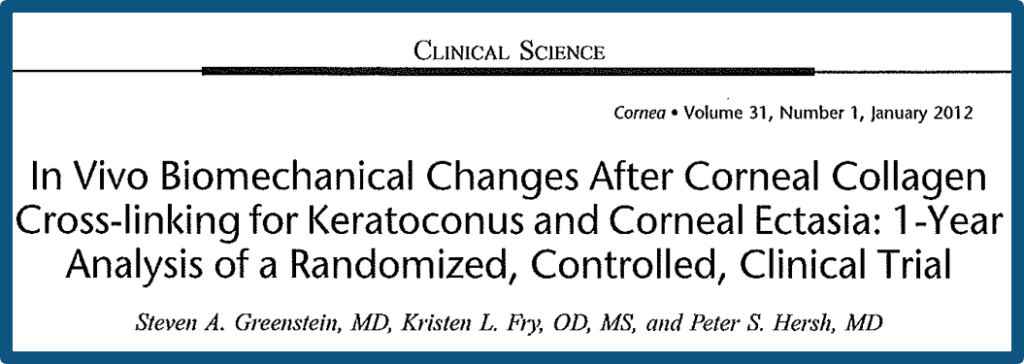
Purpose: To investigate the in vivo, corneal, biomechanical changes after corneal collagen cross-linking (CXL) using the Ocular Response Analyzer (ORA) in patients with keratoconus and post-laser in situ keratomileuses (LASIK) ectasia.
Conclusions: Despite an increase in CRF at one month, there were no statistically significant changes in CH and CRF measurements 1 year after CXL. The development of other in vivo biomechanical metrics would aid in evaluating the corneal response to CXL.
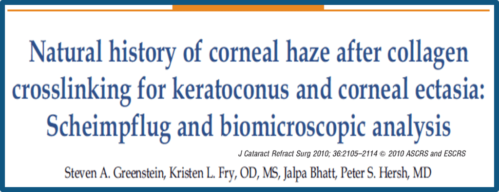
Purpose: To determine the natural history of collagen cross-linking (CXL)–associated corneal haze measured by Scheimpflug imagery and slit-lamp biomicroscopy in patients with keratoconus or ectasia after laser in situ keratomileuses.
Conclusions: The time course of corneal haze after CXL was objectively quantified; it was greatest at 1 month, plateaued at 3 months, and was significantly decreased between 3 months and12 months. Changes in haze did not correlate with postoperative clinical outcomes.
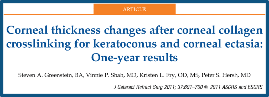
Purpose: To determine the changes in corneal thickness overtime after corneal collagen crosslinking(CXL) for keratoconus and corneal ectasia.
Conclusions: After CXL, the cornea thins and then recovers toward baseline thickness. The cause and implications of corneal thickness changes after CXL remain to be elucidated.
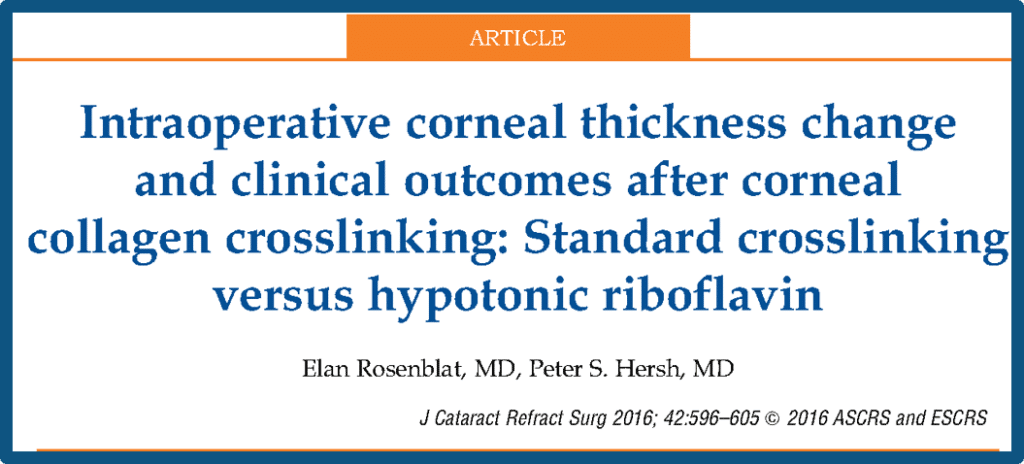
Purpose: To determine intraoperative changes in corneal thickness and outcomes of corneal collagen crosslinking (CXL) using 2 intraoperative regimens: riboflavin–dextran or hypotonic riboflavin.
Conclusions: The cornea thinned substantially during the CXL procedure. The use of hypotonic riboflavin rather than riboflavin–dextran during UVA administration decreased the amount of corneal thinning during the procedure by 30%, from 128 mm to 90 mm.
However, there were no significant differences in clinical efficacy or changes in ECC or function between groups postoperatively. In general, corneal thinning during CXL did not seem to compromise the safety of the endothelium.
