Topography-Guided PRK, Collagen Crosslinking, and Phakic IOL Implantation in Keratoconus
A 38-year-old man had been diagnosed with keratoconus 5 years before the presentation and had been stable by history. On examination, uncorrected distance visual acuity (UDVA) was counts fingers in both eyes. Corrected distance visual acuity (CDVA) was 20/20 in the right eye with a manifest refraction of -9.50-1.25 x 060 and 20/20 in the left eye with -8.00-3.50 x100.
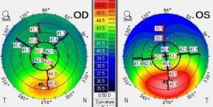
Corneal topography showed a mild keratoconus pattern in the right eye with 45.7 D maximum K and a more notable irregularity in left eye with 49.2 D maximum K. Inferior-Superior topography difference at the 6 mm zone on the axis of maximum K was 9.5D in the left eye as seen below.
TG-PRK combined with corneal collagen crosslinking was performed on the left eye. The goal was to decrease astigmatism and topographic abnormalities in preparation for ICL implantation.
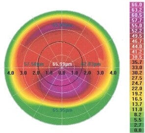
Data was imported to the Wavelight laser from repeated and reliable topography measurements. The laser pattern of the TG-PRK is seen below. The redder areas are regions where more tissue is removed and correspond to the high (red) areas of the patient’s corneal topography.
After the laser procedure, collagen crosslinking was performed. Riboflavin 0.1% was administered topically to achieve complete stromal saturation and then the cornea was aligned and exposed to UV-A (365nm) light.
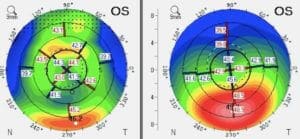
Postoperatively, the topography map showed flattening of the inferior cone and smoothing of the central optical zone. Inferior-Superior topography difference at the 6 mm zone on the axis of maximum cone height had decreased to 4.5D, indicating a 5D improvement in corneal symmetry.
The actual keratoconic cone height had decreased to 47.0 D. Glasses’ corrected vision was 20/20 with very high nearsightedness of -11.25 sphere.
Right: Before TG-PRK
The patient underwent uncomplicated placement of a phakic IOL in both eyes 3 months after the PRK/CXL procedure.
Implanted lens powers were -11.00 D in the right eye and -12.0 D in the left eye. Three months after ICL surgery, vision without glasses or contact lenses was 20/20 in the right eye and 20/16 in the left eye.

