The Annual CLEI Education
Refractive Surgery and Keratoconus Symposium
December 10 – 11, 2022
Thank you for attending our program. We are currently planning for the 2023 symposium.
FOR ALL CE CERTIFICATE-RELATED QUESTIONS PLEASE EMAIL:
Maureen Trusky : maureen@mo.events
6 COPE approved credits
Complimentary registration for all attendees
CE certificates will be delivered 6-12 weeks after the program.
About the Program
Our two-day symposium will provide education about all aspects of refractive surgery and keratoconus management.
Day one will deliver 3 CE hours on refractive surgery management.
Day two will deliver 3 CE hours on keratoconus management.
All CE hours will be broadcast live to a national and international audience, no recordings will be available for later viewing.
Register for the 2022 Symposium

Day 1: Refractive Surgery Management
Saturday, December 10, 2022
3 COPE Credits
- Refractive Surgery Diagnostics 9:30 am – 10:30 am; 1 hour CE
- Sponsor Rapid Fire 10:30 am – 11:00 am; Non CE
- Corneal Refractive Surgery 11:00 am – 12:00 pm; 1 hour CE
- Sponsor Presentations 12:00 pm – 12:30 pm; Non CE
- Lens Based Refractive Surgery 12:30 pm – 1:30 pm; 1 hour CE:

Day 2: Keratoconus Management
Sunday, December 11, 2022
3 COPE Credits
- Crosslinking for Keratoconus 9:30 am – 10:30 am; 1 hour CE
- Sponsor Presentations 10:30 am – 11:00 am; Non CE
- Surgical Options for Keratoconus 11:00 am – 12:00 pm; 1 hour CE
- Sponsor Presentations 12:00 pm – 12:30 pm
- Specialty Contact Lenses for Keratoconus 12:30pm – 1:30pm; 1 hour CE
Program Directors and Presenters

Steven A. Greenstein, MD
Cornea and Refractive Surgery
Dr. Greenstein is the director of cornea and refractive surgery at the Cornea and Laser Eye Institute. He graduated from New York University and received his M.D. from the Albert Einstein College of Medicine where he graduated with a special distinction in clinical research. Dr. Greenstein completed his Ophthalmology Residency training at Rutgers New Jersey Medical School. While at Rutgers, Dr. Greenstein trained under Dr. Peter Hersh. He completed a Cornea, Refractive, and External Disease Fellowship at Massachusetts Eye and Ear Infirmary at Harvard Medical School. He is an Assistant Clinical Professor at Rutgers New Jersey Medical School and an associate team ophthalmologist for the New York Jets. He specializes in keratoconus, corneal, and refractive surgery. His research interests include surgical keratoconus treatment and novel corneal surgery techniques. Dr. Greenstein is also involved in several clinical research studies designed to evaluate the safety and efficacy of new therapies and methods for keratoconus and refractive procedures. He has published numerous articles in prestigious medical journals; many are considered landmark papers out of the US on crosslinking. In addition to publications, he has presented at multiple scientific meetings on research related to keratoconus. He has co-authored several book chapters on corneal collagen crosslinking for keratoconus and corneal ectasia.- Peter S. Hersh, MD, FACSCornea and Refractive SurgeryDr. Hersh is the director of the research division at the Cornea and Laser Eye Institute. He graduated from Princeton University and received his M.D. from Johns Hopkins Medical School. He completed his ophthalmology residency and Cornea fellowship at the Massachusetts Eye and Ear Infirmary at Harvard Medical School. Dr. Hersh remained on the full-time faculty at Harvard for several years. In 1995, he founded the Cornea and Laser Eye Institute and, in 2002, the CLEI Center for Keratoconus. He is a Clinical Professor of Ophthalmology and Director of the Cornea and Refractive Surgery Division at Rutgers New Jersey Medical School. Additionally, he is a Visiting Research Collaborator at Princeton University. He is an internationally recognized expert in cornea and refractive surgery. He served as a principal investigator, lead author, or medical monitor for the FDA approval of landmark procedures and treatments related to refractive surgery, keratoconus, and corneal disease. Notably, the FDA approval of laser eye surgery to treat nearsightedness and corneal crosslinking for keratoconus and ectasia. He has published four textbooks, more than 100 research articles, and multiple book chapters. He was elected to the American Ophthalmological Society and awarded the Senior Honor Award from the American Academy of Ophthalmology. He is also a Life Fellow of AAO. He has trained over a thousand eye surgeons in excimer laser refractive surgery techniques. He is a past recipient of the Teacher of the Year award bestowed by the Harvard Medical School Department of Ophthalmology. As a result of his philanthropic work in several international projects to bring education and eye care to developing countries, he was awarded a Paul Harris Fellowship from Rotary International.
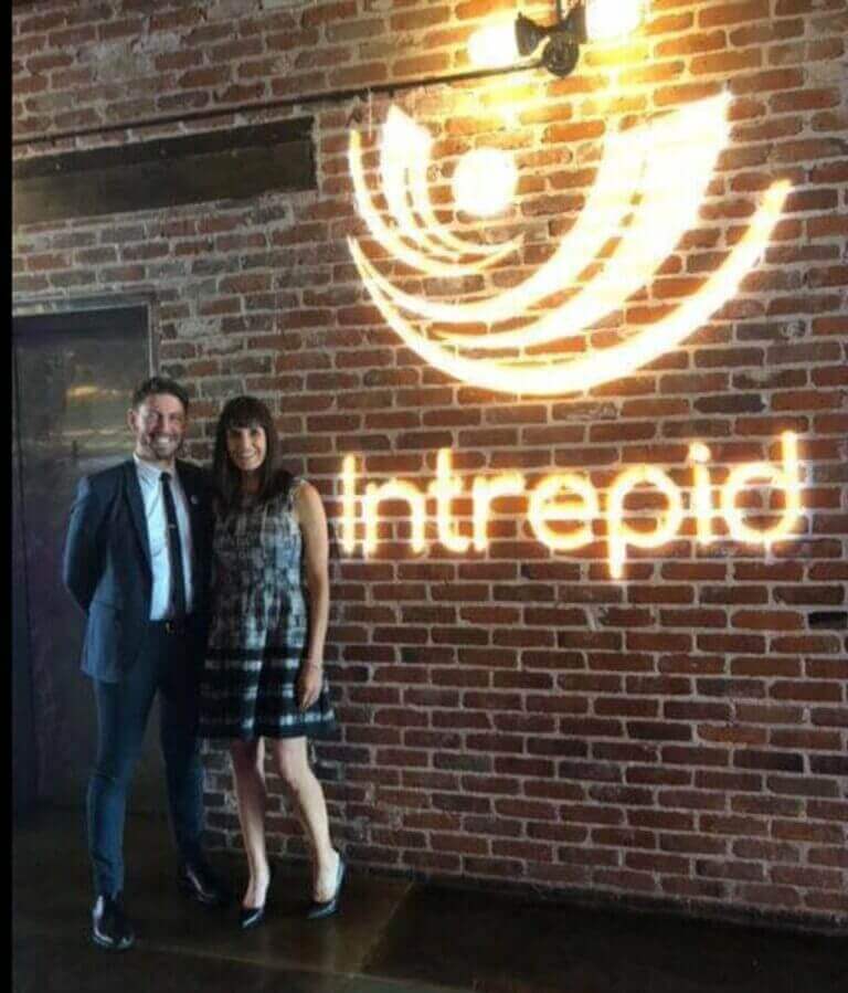
- John D. Gelles, OD, FAAO, FIAOMC, FCLSA, FSLS, FBCLASpecialty Contact Lens and PROSE ProviderDr. Gelles s the director of the specialty contact lens division at CLEI. He graduated from the University of Oregon and received his O.D. from the Pennsylvania College of Optometry. He went on to complete a Cornea Fellowship under Dr. Peter Hersh. He is an adjunct clinical professor for the New England College of Optometry and the State University of New York College of Optometry. Additionally, he is an Assistant Clinical Professor at Rutgers New Jersey Medical School. He is a PROSE clinical fellow and a fellow of the American Academy of Optometry, Scleral Lens Society, Contact Lens Society of America, British Contact Lens Association, and the International Academy of Orthokeratology and Myopia Control. In addition, he is a board member of the Contact Lens Society of America, an executive board member and education chair of the International Keratoconus Academy, an advisory board member for the Gas Permeable Lens Institute, the education chair of the Intrepid Eye Society, the chair of Refractive Surgery Alliance’s collaborative care section and serves on the American Academy of Optometry’s innovations council.He is a clinical investigator for multiple keratoconus and specialty contact lens-related clinical trials and has produced several journal articles. In addition, he regularly lectures nationally and internationally on specialty contact lenses and keratoconus and is a frequent author in several professional publications.

 Peter S. Hersh, MD, FACSCornea and Refractive SurgeryDr. Hersh is the director of the research division at the Cornea and Laser Eye Institute. He graduated from Princeton University and received his M.D. from Johns Hopkins Medical School. He completed his ophthalmology residency and Cornea fellowship at the Massachusetts Eye and Ear Infirmary at Harvard Medical School. Dr. Hersh remained on the full-time faculty at Harvard for several years. In 1995, he founded the Cornea and Laser Eye Institute and, in 2002, the CLEI Center for Keratoconus. He is a Clinical Professor of Ophthalmology and Director of the Cornea and Refractive Surgery Division at Rutgers New Jersey Medical School. Additionally, he is a Visiting Research Collaborator at Princeton University. He is an internationally recognized expert in cornea and refractive surgery. He served as a principal investigator, lead author, or medical monitor for the FDA approval of landmark procedures and treatments related to refractive surgery, keratoconus, and corneal disease. Notably, the FDA approval of laser eye surgery to treat nearsightedness and corneal crosslinking for keratoconus and ectasia. He has published four textbooks, more than 100 research articles, and multiple book chapters. He was elected to the American Ophthalmological Society and awarded the Senior Honor Award from the American Academy of Ophthalmology. He is also a Life Fellow of AAO. He has trained over a thousand eye surgeons in excimer laser refractive surgery techniques. He is a past recipient of the Teacher of the Year award bestowed by the Harvard Medical School Department of Ophthalmology. As a result of his philanthropic work in several international projects to bring education and eye care to developing countries, he was awarded a Paul Harris Fellowship from Rotary International.
 John D. Gelles, OD, FAAO, FIAOMC, FCLSA, FSLS, FBCLASpecialty Contact Lens and PROSE ProviderDr. Gelles s the director of the specialty contact lens division at CLEI. He graduated from the University of Oregon and received his O.D. from the Pennsylvania College of Optometry. He went on to complete a Cornea Fellowship under Dr. Peter Hersh. He is an adjunct clinical professor for the New England College of Optometry and the State University of New York College of Optometry. Additionally, he is an Assistant Clinical Professor at Rutgers New Jersey Medical School. He is a PROSE clinical fellow and a fellow of the American Academy of Optometry, Scleral Lens Society, Contact Lens Society of America, British Contact Lens Association, and the International Academy of Orthokeratology and Myopia Control. In addition, he is a board member of the Contact Lens Society of America, an executive board member and education chair of the International Keratoconus Academy, an advisory board member for the Gas Permeable Lens Institute, the education chair of the Intrepid Eye Society, the chair of Refractive Surgery Alliance’s collaborative care section and serves on the American Academy of Optometry’s innovations council.He is a clinical investigator for multiple keratoconus and specialty contact lens-related clinical trials and has produced several journal articles. In addition, he regularly lectures nationally and internationally on specialty contact lenses and keratoconus and is a frequent author in several professional publications.
This symposium is made possible with funding from the following industry supporters.


Please visit our friends for more educational resources


Continuing Education Event!
On November 20 – 21, 2021, The CLEI Center for Keratoconus held a complimentary 12 COPE and 6 CME Approved Course.
This symposium educated on all aspecs of refractive surgery and keratoconus management.
We plan on offering more Continuing Education Meetings for physicians in the future, so please check back for more information or e-mail info@www.vision-institute.com.
14th ANNUAL CLEI CENTER FOR KERATOCONUS PATIENT SEMINAR

On March 9, 2019, The CLEI Center for Keratoconus, a division of The Cornea and Laser Eye Institute-Hersh Vision Group held our 14th Annual Keratoconus Patient Educational Seminar at the Glenpointe Marriott Hotel in Teaneck, New Jersey.
For all who attended, the seminar was an interactive session between the participating doctors and attendees. Presentations covered the entire gamut of keratoconus from diagnosis to new treatments currently under investigation.
Dr. John Gelles, head of our Contact Lens Division started off the seminar discussing diagnosis and cause of keratoconus. He later jumped into his specialty, which is a contact lens fitting for keratoconic eyes.
At The CLEI Center for Keratoconus, Dr. Gelles fits patients with a number of different lenses, from custom soft lenses, to RGP lenses, to scleral lenses, and even the EyePrintPRO device which is a type of lens that is custom made to fit your eye.
He has achieved success, even in patients who have failed other contact lens modalities in the past. Dr. Gelles works very closely with contact lens companies with the design of new lenses to better fit the KC patient.
Dr. Peter Hersh, M.D., founder of The Cornea and Laser Eye Institute and The CLEI Center for Keratoconus lectured on new surgical strategies for keratoconus. Corneal Collagen Crosslinking (CXL), Intacs, Conductive Keratoplasty, corneal inlays, and laser procedures to rehabilitate the cornea in select KC patients, were reviewed.
Topography-guided PRK (TG-PRK) was reviewed in detail by both Dr. Hersh and Dr. Greenstein. TG-PRK is a laser procedure that may improve visual function in select keratoconus patients. With technology similar to LASIK, TG-PRK can be used in patients with keratoconus to reduce corneal optical irregularities which causes the static and decreased visual function in the eye.
The goal of this procedure is to improve the corneal shape in order to improve visual quality with glasses or contact lens fittings. CXL may also be performed concurrently, to strengthen the cornea and obtain stability. Dr. Hersh was the medical monitor of Avedro and played an important part in the FDA meeting to officially establish CXL as an FDA approved method of treatment for both keratoconus and corneal ectasia.
Dr. Steven Greenstein, M.D., corneal surgeon and keratoconus specialist, discussed the candidacy of patients for corneal collagen crosslinking (CXL). Like most procedures, it is only beneficial to you if you are a good candidate. If your cornea is below a certain micron or you have deep corneal scarring, you may not be a candidate.
Typically, patients with keratoconus tend to stabilize sometime in their 40’s – 50’s. If you have been stable for some time, CXL may not be needed in your case. At The CLEI Center for Keratoconus, we are performing a number of CXL procedures, catered to each patient.
Dr. Greenstein also discussed laser-assisted corneal transplantation (IEK) and deep lamellar keratoplasty (DALK). These are newer methods of corneal transplantation that may be beneficial in keratoconus. IEK uses a femtosecond laser to prepare both the recipient and donor cornea to match specifications. A zig-zag edge design may be made with the laser to improve graft-host fit.
Dr. David Chu, the founder of the Metropolitan Eye Research and Surgery Institute, brought his knowledge of ocular dry eye to the seminar. Dr. Chu is an international key opinion leader in areas of uveitis and ocular immunology and we are very fortunate that he is able to join us each year to discuss ocular dry eye and allergy in keratoconus patients.
Although not the only option for patients with keratoconus, corneal transplantation is still a great option if you have keratoconus, based on your doctor’s recommendation. Dr. William Constad, Partner Physician at Hudson Eye Physicians joined our physicians and discussed corneal transplantation and eye banking.
Dr. Constad, who is Director of the New Jersey Eye Bank, has a vast knowledge of the corneal tissue process. Unlike other countries, we are very lucky in the United States that more corneas are available, and unlike other organ transplants, there is typically not a waiting period for corneal tissue, should you need a transplant.
Our annual seminar is always well received and enjoyed by all participants. Each year our seminar grows and we try to improve upon topics each year. Such seminars, with the support of the National Keratoconus Foundation and other leaders in the field who join our meeting, are meant to be educational and to keep those with keratoconus, as well as their family and friends, up to date with the latest advances on the disease.
For information about attending this year’s seminar please call us at 201-692-9434 or email info@www.vision-institute.com.
The doctors of the Cornea and Laser Eye Institute, Dr. Peter Hersh, Dr. Steven Greenstein, Dr. John Gelles and Dr. David Chu, are renowned experts in their respective fields. They are frequently invited to symposia around the world to present their research and share their expertise.
Intrepid Eye Society Presents – Webinar
December 2020
Dr. Gelles presented “Innovations and Optimization of Scleral Lenses”. He and his co-presenter Dr. Melissa Barnett reviewed the most recent and upcoming innovations in scleral lens care covering everything from new scanning devices, wavefront guided optics, and future augmented reality scleral lenses. Dr. Gelles presented his early data from CLEI on wavefront guided scleral lenses, impression based scleral lenses, and scan based scleral lenses.
Indiana University – Webinar
December 2020
Dr. David S. Chu and Dr. John D. Gelles were invited to present to the optometry students at Indiana University. They presented “Connecting the Dots: Uveitis, Immunology and OSD”. Dr. Chu gave an extensive over view of uveitis and immunologic conditions as well as reviewed infusion treatments for uveitis. Dr. Gelles followed up with immunologic conditions affecting the ocular surface and their topical management.
Nova Scotia Association of Optometrists – Virtual Meeting
November 2020
Dr. John Gelles was invited to speak at the NSAO meeting this past week. He presented 2 courses “Contemporary Keratoconus Management” and “Perioperative Management of Cataract Surgery” with co-presenter Dr. Daniel Epshtein, which stressed the importance of reviewing ocular surface disease and dry eye disease prior to surgery to optimize outcomes.
World Keratoconus Day Symposium – Webinar
November 2020
World Keratoconus Day is very important to us here at the Cornea and Laser Eye Institute’s CLEI Center for Keratoconus. This year, Dr. Peter Hersh, Dr. Steven Greenstein, and Dr. John Gelles presented “Modern Management of Keratoconus”. This 2 hour course covered demographics, pathophysiology, associated disease, genetics, diagnostics, cross linking, surgical procedures, and contact lenses for keratoconus.
Refractive Surgery Alliance – Webinar
November 2020
As part of the RSA’s monthly educational series, Dr. Steven Greenstein presented “Corneal Refractive Surgery” with co-panelist Dr. Rex Hamilton. Dr. Greenstein reviewed PRK and LASIK surgery, highlighting nuances in procedure choice and selection of candidates. Dr. John Gelles served as moderator for this lecture.
American Academy of Optometry – Virtual Meeting
October 2020
This years Academy meeting was held virtually due to the COVID-19 pandemic. Dr. John Gelles was honored to be invited to present 2 courses, “Rapid Fire: Diagnostic and Management Strategies for Neurotrophic Keratitis” with co-presenters Dr. Melissa Barnett, Dr. Jennifer Harthan, and Dr. Chandra Mickles, as well as “Solving the Puzzle of Genetics for the Early Treatment of Keratoconus” with co-presenter Dr. Loretta Szczotka-Flynn.
EyeCare Partners Symposium – Virtual Meeting
October 2020
Dr. John Gelles was invited to teach a course for the EyeCare Partners Symposium. Dr. Gelles lectured on “Corneal Collagen Crosslinking: A Clinically Relevant Guide to CXL” aided at teaching his colleagues about crosslinking management for patients with keratoconus and corneal ectasia.
BostonSight Cornea Contact Lens Resident FitAcademy – Virtual Meeting
September 2020
As this year’s Cornea and Contact Lens residents continue to settle into their respective residency programs, BostonSight hosted their annual FitAcademy meeting. This intensive educational program was held virtually this year. Dr. John Gelles, a BostonSight PROSE provider, was invited to present a course titled “Scleral Lens Management of Ectasia and Irregular Astigmatism”. He and his co-instructor, Dr. Stephanie Ramdass, discussed scleral lens applications for keratoconus, pellucid marginal degeneration, post corneal surgery ectasia, and terrien’s marginal degeneration.
Refractive Surgery Alliance – Webinar
September 2020
As part of the RSA’s monthly educational series, Dr. Steven Greenstein and Dr. John Gelles presented “Refractive Surgery Diagnostics” with co-instructor Dr. Aaron Waite. Dr. Greenstein and Dr. Gelles discussed the various technologies and their importance in determining candidacy for various forms of refractive surgery.
Richens Eye Symposium – Webinar
September 2020
Dr. John Gelles was invited to teach a course for the Richens Eye Symposium. Dr. Gelles lectured on “Corneal Collagen Crosslinking: A Clinically Relevant Guide to CXL”. This course aided in teaching his colleagues about crosslinking management for patients with keratoconus and corneal ectasia.
GPLI Cornea and Contact Lens Residents Symposium – Virtual Meeting
August 2020
As this year’s GPLI Cornea and Contact Lens Residents Symposium, Dr. John Gelles was invited to teach this years residents. He was part of the “Irregular Cornea Workshop” with co-instructors Dr. Rob Ensley, Dr. Chad Rosen, Dr. Nick Gidosh and Dr. Jason Jedlicka and the “Scleral and Hybrid Workshop” with co-instructors Dr. Rob Ensley, Dr. Chad Rosen, Dr. Stephanie Woo, and Dr. Jason Jedlicka.
Intrepid Eye Society Presents – Webinar
July 2020
At this month’s Intrepid Eye Society peer education webinar, Dr. John Gelles presented “Management Strategies for Severe OSD: From Amnion to Scleral Lenses” with co-presenters Dr. Scott Hauswirth, Dr. Bita Asghari, and Dr. Jacob Lang. This course taught colleague about complex ocular surface disease and treatment options ranging from topical medications, PROSE treatment, therapeutic contact lens wear, immunologic therapy, biologic therapy, and surgical intervention.
Contact Lens Society of South Africa – Webinar
July 2020
Dr. John Gelles was honored to be invited to present to the CLSSA. Dr. Gelles presented on “Current and Future Contact Lens Innovation: From surface based lens design and HOA correcting optics, to displays, photonics and drugs” with co-presenter Dr. Christine Sindt.
Intrepid Eye Society Presents – Webinar
June 2020
At this month’s Intrepid Eye Society peer education webinar, Dr. John Gelles presented “Smartphone and Tablet Based Slit Lamp Photography: Techniques and Considerations for Anterior Segment Imaging”. This course taught advanced techniques for capturing subtle corneal pathology and included live virtual demonstration.
World Ophthalmology Congress – Virtual Meeting
June 2020
It was an honor for Dr. John Gelles to be invited to lecture at the prestigious World Ophthalmology Congress. Dr. Gelles was part of an expert panel titled “Therapeutic Contact Lenses – Where Are We in 2020?” co-panelist Dr. Sorcha Ni Dhubhghaill, Dr. Oliver Woo OD, Dr. Christina Grupcheva, and Dr. Christine Sindt. He presented a course titled “Current and Future Contact Lens Innovation: From surface based lens design and HOA correcting optics, to displays, photonics and drugs”.
Intrepid Eye Society Presents – Webinar
May 2020
At this month’s Intrepid Eye Society peer education webinar, Dr. John Gelles presented “Ocular Surface Disease and Objective Image Analytics: From TFOS DEWS II to The ASCRS Algorithm, Why it Matters for OD’s, MD’s and Collaborative Care” with co-instructor Dr. Christopher Starr. This lecture taught a practical, in depth review of the majority OSD literature and related this to the importance of objective analytics for precise, accurate, and reproducible grading of OSD severity.
Scleral Lens Education Society – Webinar
April 2020
Dr. John Gelles was invited by the Scleral Lens Education Society to teach on a very timely topic, “Telemedicine and Scleral Lenses: A Panel Discussion” with co-panelists Dr. Marcus Noyes and Dr. Clarke Newman. Telemedicine has become extremely important due to the spread of coronavirus. Patients are currently in need of care and during this pandemic it is important doctor’s know how to use telemedicine to care for their scleral lens wearing patients.
Cornea and Laser Eye Institute and CLEI Center for Keratoconus Symposium – Webinar
February 2020
Dr. Peter Hersh, Dr. Steven Greenstein and Dr. John Gelles presented a national webinar on “Topography-guided PRK for Keratoconus”. Dr. Greenstein reviewed the data from CLEI about the TGPRK procedure, Dr. Gelles reviewed data on TGPRK effects contact lens design and data from a series of contact lens fits on post TGPRK patients, and Dr. Hersh reviewed the technology behind the procedure and individual cases.
New Jersey Society of Optometric Physicians – Randolph, NJ
February 2020
Dr. Steven Greenstein and Dr. John Gelles were invited to present at the NJSOP’s meeting. Together they presented “Innovations in Keratoconus Management” where Dr. Greenstein reviewed some of the newest surgical procedures, topography guided PRK and corneal tissue inlays, and Dr. Gelles discussed the newest contact lenses, scan based scleral lenses and wavefront guided scleral lenses.
CEWire – Virtual Meeting
January 2020
Dr. John Gelles was invited back this year to speak at one of the largest virtual meetings, CEWire. This year Dr. Peter Hersh and Dr. Steven Greenstein joined Dr. Gelles to present “Contemporary Keratoconus Management”. Dr. Gelles reviewed methods for early detection of keratoconus and metrics for monitoring the disease, Dr. Greenstein reviewed methods of corneal collagen cross-linking for keratoconus and clinical data from CLEI, and Dr. Hersh reviewed surgical methods for vision improvement and Dr. Gelles wrapped up reviewing contact lens options for keratoconus. Dr. Gelles also presented “Future Keratoconus Management: A Glimpse Of What’s To Come”, which reviewed the literature to find and discuss the most exciting research on upcoming devices, surgical and nonsurgical treatments, and finally “Custom Soft Lenses for Keratoconus and Beyond” which reviewed options and methods of soft lenses fitting for keratoconus and other ectatic corneal disease.
Global Specialty Lens Symposium – Las Vegas, NV
January 2020
It was a honor for Dr. John Gelles to be invited back to lecture at the 2020 Global Specialty Lens Symposium. Dr. Gelles presented 3 course, “Keratoconus: What You Need to Know: Diagnostics, Cross-Linking, and Scleral Lenses”, “Contact Lenses for Visual Rehabilitation in Keratoconus”, and “Foundations of a Specialty Contact Lens Practice”.
Scleral Lens Education Society – Webinar
January 2020
This month Dr. John Gelles was honored to present “Corneal Collagen Crosslinking for Keratoconus” as part of the Scleral Lens Education Society’s webinar. This course reviewed corneal pathophysiology, international foundational data about the procedure, and data from the US clinical trial where CLEI was the primary clinical trial site and Dr. Hersh served as principal investigator and medical monitor.
New York Academy of Optometry Meeting – New York, NY
December 2019
Dr. Steven Greenstein and Dr. John Gelles were invited to present at the NYAO meeting. Together they presented “Keratoconus Update: Surgical and Non-Surgical Treatment” where Dr. Greenstein reviewed topography guided PRK and corneal tissue inlays and Dr. Gelles discussed scan based scleral lenses and wavefront guided scleral lenses.
Cornea and Laser Eye Institute and CLEI Center for Keratoconus Symposium – Webinar
October 2019
Dr. Peter Hersh, Dr. Steven Greenstein and Dr. John Gelles presented a national webinar on “Modern Management of Keratoconus”. Dr. Gelles review methods for early detection of keratoconus and metrics for monitoring the disease, Dr. Greenstein reviewed methods of corneal collagen cross-linking for keratoconus and clinical data from CLEI, and Dr. Hersh reviewed surgical methods for vision improvement and Dr. Gelles wrapped up reviewing contact lens options for keratoconus.
3rd World Congress of Optometry – Orlando, FL
October 2019
Following the American Academy of Optometry’s Annual Meeting, Dr. Gelles was invited to speak at the World Congress of Optometry. This international meeting happens every 4 years and brings together doctors and researchers from around the globe. Dr. Gelles presented a lecture entitled “FDA Approved Corneal Collagen Crosslinking: A Clinically Relevant Guide to CXL”. This lecture reviewed in depth the biomechanics of keratoconus, biochemistry of crosslinking, and the body of research which affects the clinical follow up of patients undergoing CXL treatment.
American Academy of Optometry Meeting – Orlando, FL
October 2019

Dr. John Gelles was delighted to serve as a panelist, along with Dr. Renee Reeder and Dr. Randall Sakamoto with Dr. Joe Barr as moderator, during the keratoconus roundtable. This roundtable was sponsored by The National Keratoconus Foundation. AAO is a great meeting to discuss KC contact lens options as a group. The experts (Dr. Gelles) chosen offered opinions, practice tips and insights from their experience managing patients with keratoconus. Additionally Dr. Gelles presented two lectures, one titled “Rapid Fire: The Next Generation in Scleral Lens Fitting” with co-presenter Dr. Brooke Messer, Dr. Sheila Morrison and Dr. Maria Walker and the other “Contact Lenses for Visual Rehabilitation in Keratoconus”.
East West Eye Conference – Cleveland, OH
October 2019
Dr. Gelles was honored to be invited to speak at the East West Eye Conference. He presented on “Contact Lenses for Visual Rehabilitation in Keratoconus” reviewing outcomes of contact lens wearers from CLEI. He was also part of a session called “Discussion on Keratoconus: Modern Management” where he lectured with colleagues Dr. Loretta Szczotka-Flynn, who taught on the genetics of keratoconus, and Dr. William Dupps, who taught on the biomechanics of keratoconus. Dr. Gelles taught on modern diagnostics and early detection of keratoconus.
Contact Lens Society of America Meeting – Orlando, FL
September 2019
At this year’s Contact Lens Society of America meeting, Dr. Gelles was invited to present two lectures and participate in several panels. He presented “The Latest and Greatest Clinical Tools to Maximize Scleral Lens Fitting” reviewing new and upcoming technology and “Custom Soft Lens Technology Indications and Contraindications”. Dr. Gelles was a panelist in both, the Contact Lens Grand Rounds: Myopia Control session with co-panelists Dr. Jason Jedlicka and Dr. David Kading, and the Contact Lens Grand Rounds: Scleral Lenses session with co-panelists Dr. Jason Jedlicka and Dr. Stephen Byrnes.
Advanced Refractive Congress – Miami, FL
July 2019
It was a honor for Dr. John Gelles to be invited back to lecture at this years Advanced Refractive Congress. Dr. Gelles presented “Contact Lens Trends in Keratoconus After CXL, Intacs, and TGPRK”. This presentation reviewed data from CLEI showing a spectrum of contact lenses used in post CXL and TGPRK eyes and finding that mainly soft and scleral designs are used in Intacs.
International Congress of Scleral Contacts – Miami, FL
July 2019
Dr. John Gelles was honored to be invited to lecture at this year’s ICSC. He served as a panelist for the “The Atypical Scleral Lens Patient” session with Dr. Ryan McKinnis and Dr. Priscilla Sotomayor. This session focused on patients that are outside of the norm for scleral lenses, such as patients that have experienced traumas, infections, or surgeries resulting in an irregular corneal shape.
Solinsky Eye Care Symposium – Hartford, CT
April 2019
Dr. John Gelles was honored to be invited to lecture at the Solinsky Eye Care Symposium. He presented “FDA Approved Corneal Collagen Crosslinking for Keratoconus”. Additionally, he lectured with Dr. Michael Cooper to present “Cornea on (Rapid) Fire”, a case based lecture reviewing a spectrum of corneal disease states and management. Finally Dr. Gelles and Dr. Cooper were joined by Dr. Susan Janik to present a “Dry Eye Workshop” reviewing everything from diagnostics to procedures for dry eye disease.
Connecticut Opticians Association Meeting – Middletown, CT
March 2019
It was a true honor for Dr. John Gelles to be invited back to speak with the COA as the primary instructor for the COA contact lens education tract. He presented 8 hours of lectures including “Keratoconus Management Grand Rounds: How would you treat this patient?”, “Contemporary Keratoconus Management Diagnose Early, Stop Progression, Rehabilitate Vision”, “Tricks and Tips for Successful Therapeutic Scleral Lens Wear in Ocular Surface Disease”, “Scleral Lens Troubleshooting”, “Custom Soft Lenses for Keratoconus and Beyond” and “Non-Surgical Visual Rehabilitation for Keratoconus Patients”.
Gas Permeable Lens Institute – Webinar
February 2019
It was an honor for Dr. John Gelles to be invited to speak as part of the GPLI education series. The lecture was titled “Technology Applications in Specialty Contact Lens Practice”. In the course Dr. Gelles highlighted new devices to aid in specialty contact lens designs and future developments to aid in monitoring corneal disease.
Connecticut Opticians Association Meeting – Middletown, CT
November 2018
Dr. John Gelles was invited to speak with the COA as the primary instructor for their contact lens tract. He presented 8 hours of lectures including “Keratoconus Management Grand Rounds: How would you treat this patient?”, “Contemporary Keratoconus Management Diagnose Early, Stop Progression, Rehabilitate Vision”, “Tricks and Tips for Successful Therapeutic Scleral Lens Wear in Ocular Surface Disease”, “Scleral Lens Troubleshooting”, “Custom Soft Lenses for Keratoconus and Beyond” and “Non-Surgical Visual Rehabilitation for Keratoconus Patients”.
Westchester, Rockland, Putnam Optometric Society Meeting – Westchester, NY
October 2018
This month Dr. John Gelles presented “Contemporary Keratoconus Management Diagnose Early, Stop Progression, Rehabilitate Vision” to Westchester, Rockland, Putnam Optometric Society. This course taught colleagues about comprehensive management of keratoconus.
Vision Expo West – Las Vegas, NV
September 2018
Dr. John Gelles was invited to present at Vision Expo West. He presented “Custom Soft Lenses for Keratoconus and Beyond” and “Keratoconus Management Grand Rounds: How would you treat this patient ?”. As well as an encore lecture and workshop from Vision Expo East, “Scleral Lens Troubleshooting” with co-presenter Dr. Woo and “Scleral Lens Workshop” with co-instructors Dr. Barnett, Dr. Chang and Dr. Newman.
Optometric Society of the City of New York Meeting – New York, NY
August 2018
This month Dr. John Gelles presented “Contemporary Keratoconus Management Diagnose Early, Stop Progression, Rehabilitate Vision” to the Optometric Society of the City of New York. This course taught doctors about comprehensive management of keratoconus.
International Congress of Scleral Contacts – Miami, FL
July 2018
Dr. John Gelles was honored to be invited to lecture at this year’s ICSC. He served as a moderator for the “Scleral Lenses Necessities” session and also gave a lecture by the same name. In the lecture he discussed everything from educational resources to equipment necessary to deliver scleral lens care.
Advanced Refractive Congress – Miami, FL
July 2018
Dr. John Gelles was honored to be invited to speak at this year’s Advanced Refractive Congress. This year he presented on “Objective Image Analytics: Automated Grading”. This lecture focused on the use of software to grade severity of various disease states and produce consistency, accuracy and repeatability to eliminate variability and bias in clinical practice, clinical trials and research.
Gas Permeable Lens Institute – Webinar
June 2018
Dr. John D. Gelles was invited to speak as part of the Gas Permeable Lens Institute’s education series. Dr. Gelles presented “Contact Lens Fitting in Keratoconus Post Surgery”. This lecture discussed data from CLEI reviewing success rate with various types of specialty lens in patients after undergoing crosslinking, Intacs, and topography guided PRK.
Annual Intrepid Eye Society Meeting – Denver, CO
June 2018
This year’s Intrepid Eye Society Meeting was held in Denver, Colorado. At the meeting, Dr. Gelles presented “Scleral Lenses for Ocular Surface Disease” with co-presenter Dr. Melissa Barnett.
Vision by Design – Orlando, FL
April 2018
Dr. Gelles was invited to speak at this year’s Vision by Design meeting to present “Contemporary Keratoconus Management: Diagnose Early, Stop Progression, Rehabilitate Vision” during the corneal disease symposium. He and his co-presenter, Dr. Andrew Morgenstern ran an educational workshop as well, teaching other doctors the nuances of keratoconus diagnosis and monitoring.
Vision Expo East – New York, NY
March 2018
This year at Vision Expo East, Dr. Gelles was invited to present “Scleral Lens Troubleshooting”. He and his co-presenter, Dr. Stephanie Woo reviewed the nuances to successful scleral lens design. He also taught a hands on workshop, “Scleral Lens Workshop” teaching other doctors how to fit scleral lens, with co-instructors Dr. Melissa Barnett, Dr. Clark Chang, and Dr. Clarke Newman.
Global Contact Lens Forum – New York, NY
March 2018
Prior to Vision Expo East, Dr. Gelles was invited to be a panelist and present at the Global Contact Lens Forum. The panel session was moderated by Dr. Louise Sclafani and Dr. Barry Eiden. Dr. Gelles presented “Scleral Lenses: Visual Rehabilitation and Therapeutic Use”.
Sonoma Eye Symposium – Sonoma, CA
February 2018
Dr. David S. Chu is the education chair and meeting founder of Sonoma Eye, a meeting dedicated to ocular inflammatory and immunologic disease. At the meeting Dr. Chu presented on “New Biologic Treatments for Uveitis” and Dr. Gelles presented on “Scleral Lenses for Ocular Surface Disease”.
Global Specialty Lens Symposium – Las Vegas, NV
January 2018
Dr. Gelles was recently invited to present at this year’s Global Specialty Lens Symposium. He presented case based lecture title “Keratoconus Management Grand Rounds: How would you treat this patient?”. The presentation reviewed modern diagnostics and treatment approaches to keratoconus.
Scleral Lens Education Society – Webinar
December 2017
Dr. Gelles lectured as part of the Scleral Lens Education Society’s education series. He presented “Tricks and Tips for Successful Therapeutic Scleral Lens Wear in Ocular Surface Disease” with co-presenter Dr. Melissa Barnett. The lecture discuss strategies employed by him and Dr. Barnett to facilitate success in therapeutic scleral lens wear.
Cornea, Contact Lens & Contemporary Vision Care Symposium – Houston, TX
December 2017
Dr. Gelles was invited to present at this year’s Cornea, Contact Lens & Contemporary Vision Care Symposium. He presented “Keratoconus: Early Detection and Contemporary Monitoring” where he discussed established guidelines for early diagnosis and monitoring and proposed new diagnostic methods.
International Forum for Scleral Lens Research – Houston, TX
December 2017
Dr. Gelles was invited to present at this years IFSLR, he presented “IOP and Scleral Lens: Factors, Limitations, and Novel Approaches” exploring concepts in scleral lens wear and it’s impact on biomechanics and intraocular pressure and proposed various techniques and methods of evaluating pressure changes during lens wear.
America Academy of Optometry Meeting – Chicago, IL
October 2017
Dr. Gelles was invited to present at this year’s American Academy of Optometry Meeting. He present two lectures, “Scleral Lens Virtual Workshop: You Choose the Parameters” with co-presenter Dr. Jedlicka and “Optimizing Ocular Health in Scleral Lens Fitting: Adopt the Minimalist Approach” with co-presenters Dr. Jason Jedlicka, Dr. Langis Michaud, and Dr. Daniel Brazeau.
Contact Lens Society of America Meeting – Orlando, FL
September 2017
Dr. Gelles was invited to present at this year’s Contact Lens Society of America meeting. He presented on “Tricks and Tips for Successful Therapeutic Scleral Lens Wear in Ocular Surface Disease” and “Non-Surgical Visual Rehabilitation for Keratoconus Patients”. During this meeting he was honored with the Keith Harrison Award.
American Academy of Ophthalmology – Webinar
August 2017
Dr. Peter S. Hersh, along with Dr. William B. Trattler, presented a live webinar for the American Academy of Ophthalmology on Corneal Collagen Crosslinking. The goals were to gain an understanding of the background and development of CXL as well as determining whether CXL is the most appropriate treatment for the patient.
Advanced Refractive Congress – Miami, FL
July 2017
Dr. John D. Gelles was invited to lecture at this years Advanced Refractive Congress. His lecture “Intacs in the Age of Crosslinking: A Clinical Review” reviewed the use of Intacs combined with CXL, an results from the prospective clinical trial at CLEI where patients were randomized to either sequential or concurrent CXL and Intacs. He also reviewed CLEI data on complications and removals of Intacs, discuss these rare events.
International Congress of Scleral Contacts – Miami, FL
July 2017
Dr. Gelles was invited to lecture at The International Congress of Scleral Contacts meeting this past July. During his talk, “Therapeutic Scleral Lenses: Stain WITH and WITHOUT Pain”, he discussed therapeutic scleral lens wear for patients with complex ocular surface disease.
The Cornea and Laser Eye Institute – CLEI Center for Keratoconus Symposium – Teaneck, NJ
May 2017
Dr. Peter Hersh and Dr. John Gelles hosted colleagues at an educational event on keratoconus. Together they discussed Corneal Collagen Crosslinking and surgical and non-surgical strategies for comprehensive keratoconus management over this 3 hour event.
New York Academy of Optometry Meeting – New York, NY
March 2017
Dr. John D. Gelles and Dr. Peter S. Hersh were invited to speak at the NYAO meeting. Dr. Gelles presented on “Non-Surgical Visual Rehabilitation for Keratoconus” discussing various specialty contact lens options for keratoconus and “Diagnostics and Early Detection of Keratoconus” reviewing established metrics and upcoming technologies. Dr. Hersh presented on “FDA Approved Corneal Collagen Cross-linking for Progressive Keratoconus & Corneal Ectasia” discussing the procedure and results of the FDA clinical trial for CXL where he served as principal investigator and medical monitor.
Vision Monday Global Leadership Summit – New York, NY
March 2017

It was a honor for Dr. John Gelles to be invited to present at this year’s Vision Monday Global Leadership Summit. This year’s summit was titled “A-Eye – Artificial Intelligence: Supercharging Knowledge and Decision Making”. Dr. Gelles presented as part of a panel on Artificial Intelligence, Machine Learning and Telemedicine. He presented on “Telemedicine Solutions for Eye Care” with co-panels, Dr. Pearse A. Keane, Dr. Paul Karpecki, and Dr. Kovin Naidoo.
Gas Permeable Lens Institute – Webinar
January 2017
Dr. John Gelles was invited to speak as part of the GPLI education series. The lecture was titled “Scleral Lens Fitting to Optimize Corneal Physiology” with co-presenter Dr. Michaud. They discussed challenges related to scleral lens wear and methods to improve physiology in compromised corneas.
International Forum for Scleral Lens Research – Houston, TX
December 2016
Dr. John Gelles was invited to be a panelist at the first International Forum for Scleral Lens Research meeting. He discussed the role of oxygen permeability of scleral contact lens system.
American Academy of Ophthalmology, Annual Meeting – Chicago, IL
October 2016
Dr. Peter Hersh was invited to participate as a speaker at the Avedro North Lecture Series at AAO. He discussed the U.S. FDA Approved Cross-Linking Keratoconus and Corneal Ectasia Data. Dr. Hersh was medical monitor of the clinical trials which led to recent FDA approval.
International Congress of Scleral Contacts 2016 – Miami, FL
July 2016
Dr. John Gelles was invited to lecture at The International Congress of Scleral Contacts meeting this past July. During his talk, “EyePrintPRO for Bilateral Proud Grafts”, he discussed scleral lens complications, scleral lenses and corneal transplants, as well as EyePrintPRO for bilateral proud grafts when all other options failed. Dr. Gelles is among a group of less than 25 trained providers worldwide who fits patients with the EyePrintPRO device. We are excited to be able to offer this technology to our patients!
First Annual Intrepid Eye Society Meeting – Boston, MA
June 2016
Dr. John Gelles, a founding member of the Intrepid Eye Society, presented, “Combined Procedures Keratoconus” and “Telemedicine of Eyecare” to a group of optometrists. These lectures covered current and future procedures for keratoconus, current and future research at The Cornea and Laser Eye Institute, as well as the role of telemedicine in eyecare.
The Intrepid Eye Society is a diverse group of emerging thought leaders in optometry. Their initiatives include advancement of optometry through innovative thought sharing on topics related to future medical therapeutics, diagnostics, practice development, research and development, and collaborative care and ophthalmology.
New York Academy of Optometry Meeting – New York, NY
June 2016
Dr. John Gelles was invited to discuss Corneal Collagen Crosslinking and Scleral Lenses at the NYAO meeting. The lectures were titled, “FDA Approved Corneal Collagen Cross-linking for Progressive Keratoconus” and “Fitting Philosophies for Oxygen Delivery and Successful Scleral Lens Wear in Diseased Corneas”.
Dr. Gelles is the Director of the Specialty Contact Lens Division here at The Cornea and Laser Eye Institute – CLEI Center for Keratoconus.
The Cornea and Laser Eye Institute – CLEI Center for Keratoconus Symposium – Teaneck, NJ
May 2016
Dr. Peter Hersh and Dr. John Gelles invited local optometrists to an educational event on Corneal Collagen Crosslinking. Corneal Collagen Crosslinking was recently FDA approved and granted to Avedro, Inc. for Photrexa Viscous, Photrexa and the KXL System. Dr. Hersh was medical monitor of the clinical trial which led to this approval. Dr. Hersh discussed FDA approval for patients with progressive keratoconus while Dr. Gelles discussed how to successfully comanage patients who are having crosslinking performed.
If you are a physician interested in future continuing educational events at The Cornea and Laser Eye Institute, please contact Stacey Lazar at slazar@www.vision-institute.com.
The Association for Research in Vision and Ophthalmology (ARVO)
2011 Annual Meeting – May 1 – 5, 2011
Title: Two Year Outcomes After Corneal Collagen Crosslinking For Keratoconus and Ectasia
Session Title: Corneal Biomechanics, Keratoconus and Collagen Cross Linking
Paper:
Purpose: To investigate clinical outcomes two years after corneal collagen crosslinking (CXL) for keratoconus (KC) and ectasia.
Methods: 50 eyes (38 patients) underwent CXL for keratoconus (n=34) or ectasia (n= 16) in a prospective, randomized controlled trial. CXL was performed with a UVX system (Peschke Meditrade GmbH) using standard technique. Clinical outcomes including uncorrected visual acuity (UCVA), best corrected visual acuity (BCVA), maximum keratometry (Kmax), CXL associated corneal haze, and corneal thickness were assessed and analyzed at baseline, 1 month, 3 month, 6 month, 1 year, and 2 year follow up visits. Corneal haze was measured by Scheimpflug densitometry.
Results: In the entire cohort, mean UCVA preoperatively was 20/151 (logMAR 0.88±0.35) and improved at 2 years to 20/136 (logMAR 0.83±0.38, p=0.2). BCVA was significantly improved from 20/46 (logMAR 0.36±0.24) to 20/35 (logMAR 0.24±0.19) (p<0.001). On topography, Kmax flattened significantly by 2.0±4.7D (p=0.004). Pachymetry values at 2 years were not significantly different from baseline (p=0.3). Corneal haze remained slightly increased at 2 years compared to baseline (+1.33±3.8, p=0.02). Between 1 and 2 years, the change in UCVA (change=-0.02±0.30, p=0.7), BCVA (change=-0.04±0.16, p=0.08), and Kmax (change=-0.05±2.9D, p=0.9) were not statistically significant. Mean corneal haze (change=-1.17±4.7), and pachymetry (change=+3.4±21.8μm), both continued to return toward baseline measurements, but these changes failed to reach statistical significance as well (phaze=0.09, ppachymetry=0.3). A similar clinical course was found in the analysis of stratified KC and ectasia subgroups.
Conclusions: At 2 years, CXL was effective at improving visual acuity and topographic outcomes. Clinical outcomes remained stable between 1 and 2 years after CXL.
Title: Corneal Thickness Effects using Riboflavin/Dextran versus Hypotonic Riboflavin during Corneal Collagen Crosslinking
Session Title: Corneal Biomechanics Keratoconus and UV Crosslinking
Paper:
Purpose: To investigate the use of Riboflavin 0.1% in 20% dextran solution versus its hypotonic formulation during ultraviolet light exposure in corneal collagen crosslinking (CXL) for keratoconus (KC) and ectasia.
Methods: In a prospective, controlled clinical trial, 21 eyes underwent CXL for KC or ectasia (14 and 7 eyes respectively). CXL was performed using the UVX system (Peschke Meditrade GmbH). All eyes underwent pre-treatment with Riboflavin 0.1% in 20% dextran solution every 2 minutes for 30 minutes. If the cornea was <400 microns thick, then hypotonic riboflavin was administered until the cornea swelled beyond 400 microns. Eyes were then randomized to receive either Riboflavin 0.1%/dextran (RIBO) or hypotonic (HYPO) riboflavin every 2 minutes for the 30 minute duration of UV exposure (UVA 365nm light for 30 minutes at an irradiance of 3mW/cm2). Corneal thickness was measured using ultrasound pachymetry (Sonogage).
Results: Mean initial corneal thickness was 435.7μm (ranging 318 to 526) and 445.4μm (ranging 327 to 532) for RIBO and HYPO eyes respectively. Following debridement, mean thickness was 392.1μm (ranging 304 to 463) for RIBO eyes and was 402.9μm (ranging 330 to 454) for HYPO eyes. After pre-treatment with riboflavin 0.1%/dextran, mean thickness was 417.2μm (ranging 323 to 535) for RIBO eyes and 417.0μm (ranging 317 to 468) for HYPO eyes. Three eyes (2 RIBO; 1 HYPO) required the use of hypotonic riboflavin to achieve the minimum corneal thickness of 400μm prior to UV irradiance. Mean corneal thinning during UV exposure was 133.8μm (SD 33.2)(p<0.0001) and 94.6μm (SD 48.0)(p=0.002) for RIBO and HYPO eyes respectively. Mean corneal thinning with UV exposure was significantly greater for RIBO eyes compared to HYPO eyes (p=0.04) At the conclusion of CXL treatment, mean thickness was 294.4μm (ranging 238 to 345) and 335.3μm (ranging 271 to 398) in RIBO and HYPO eyes respectively.
Conclusions: The use of hypotonic riboflavin appears to better maintain consistent corneal thickness during UV administration. Whether better maintenance of corneal thickness potentially may result in improved consistency, reproducibility and/or safety of the CXL procedure requires further follow-up.
Title: Corneal Collagen Crosslinking Outcomes With and Without Stromal Swelling with Hypotonic Riboflavin
Session Title: Corneal Biomechanics Keratoconus and UV Crosslinking
Paper:
Purpose: It has been suggested that hypotonic riboflavin can be used to swell the corneal stroma to a safe level in order to perform corneal collagen crosslinking (CXL). In this investigation, we compare 1 year CXL clinical outcomes, using hypotonic riboflavin for corneal swelling vs. standard riboflavin dextran solution only.
Methods: 103 eyes underwent CXL for keratoconus (n=68) or ectasia (n= 35) in a prospective, randomized controlled trial. Riboflavin (0.1% in 20% dextran T500 solution) was administered topically for 30 minutes. If the cornea was <400µm after this dosing, hypotonic riboflavin (0.1% in sterile water) was given, 1 drop every 10 seconds for 2 minute rounds, and ultrasonic pachymetry (U/S) was performed until the stroma had swollen to >400µm. Uncorrected visual acuity (UCVA), best corrected visual acuity (BCVA), maximum keratometry (Kmax), corneal haze, and thinnest pachymetry (Pthin) were compared between riboflavin groups.
Results: In the riboflavin dextran only group (n=42), UCVA (logMAR change=-.07±0.19,p=.04), BCVA (logMAR change=-.09±0.17,p<.01), and Kmax (change=-1.2±1.9D,p<.01) significantly improved, and corneal haze returned to baseline at 1 year (p=.1). Preoperative Pthin was 468.2±48.2µm, and at 1 year was slightly thinner (458.2±47.5µm,p<.01). In the hypotonic group (n=61), preoperative U/S, U/S after 30 minutes of riboflavin dextran administration, and U/S after hypotonic riboflavin administration, was 423.9±53.7µm, 340.9±45.1µm, and 413.6±11.1µm, respectively. On average, 3.1±2.2 rounds of hypotonic drops were required to swell the cornea ≥400µm. In this group, UCVA (logMAR change=-.09±0.28,p=.01), BCVA (logMAR change=-.1±0.18,p<.01), and Kmax (change=-1.9±4.0D,p<.01) significantly improved, and corneal haze remained slightly above baseline at 1 year (p<.01). Preoperative Pthin was 423.1±51.1µm, and at 1 year returned to preoperative measurement (420.9±51.7µm,p=.4). There was no significant difference between the two riboflavin groups, when comparing the changes in any of these clinical outcomes at 1 year (PUCVA =.5, PBCVA =.6, PKmax = .2, PHaze =.1, PPthin = .1).
Conclusions: Administration of hypotonic riboflavin to swell the corneal stroma does not appear to affect 1 year clinical outcomes compared to standard riboflavin/dextran alone.
Title: Simultaneous versus Sequential Intacs and Cornea Collagen Crosslinking for Keratoconus and Ectasia
Session Title: Corneal Biomechanics, Keratoconus and Collagen Cross Linking
Paper:
Purpose: To investigate the optimum timing of Intacs combined with corneal collagen crosslinking (CXL) (simultaneous vs. sequential) for keratoconus and ectasia.
Methods: In a prospective, controlled clinical trial, 31 eyes were randomized to receive Intacs followed by CXL on the same day (Sim group) (15 eyes) or Intacs followed by CXL at 3 months postoperatively (Seq group) (16 eyes). All eyes received symmetric 350 micron Intacs segments (Addition Technology Inc.) and CXL was performed using the UVX system (Peschke Meditrade GmbH). Clinical outcomes including UCVA, BCVA, MRSE, manifest cylinder, and topographic changes (Kmax) using the Pentacam were assessed pre- and postoperatively.
Results: Thirteen eyes in the Sim group and 7 eyes in the Seq group have data collected to 3 months postoperatively. For Sim eyes, mean UCVA preoperatively was 20/171 and was 20/162 three months postoperatively. Mean BCVA changed from 20/44 preoperatively to 20/47 at 3 months postoperatively. MRSE improved by 0.94D and manifest cylinder remained the unchanged. Topographically, Kmax flattened by 0.91D. All outcomes failed to reach statistical significance. Similarly, Seq eyes, showed no significant changes in UCVA and BCVA which improved from 20/175 to 20/153 and 20/36 to 20/28 at 3 months postoperatively respectively. MRSE improved by 0.68D, but failed to reach statistical significance. Manifest cylinder remained unchanged as well as topographically measured mean Kmax. No significant differences between Sim and Seq cohorts were observed.
Conclusions: Early results show no differences in Sim compared to Seq Intacs and CXL. Studies suggest that the beneficial effects of CXL are not achieved until 6 months postoperatively which indicate early healing may be confounding results. The optimal method timing of Intacs combined with CXL remains to be elucidated with further follow-up.
Title: Patient Satisfaction and Subjective Visual Function after Corneal Collagen Crosslinking
Session Title: Corneal Biomechanics Keratoconus and UV Crosslinking
Paper:
Purpose: To assess patient satisfaction and subjective visual function, using a patient questionnaire, one year after corneal collagen crosslinking (CXL) for keratoconus and corneal ectasia.
Methods: 107 eyes of 76 patients underwent CXL for keratoconus (n=71) or ectasia (n= 36) in a prospective, randomized controlled trial. Patients completed a subjective questionnaire concerning their visual symptoms, administered preoperatively and at 1 year. Patients ranked symptoms on a scale from 1 to 5 (1= none, 2= mild, 3=moderate, 4=marked, 5= severe). Reported symptoms of photophobia, difficulty night driving, difficulty reading, diplopia, fluctuations in vision, glare, halo, starburst, dryness, pain, and foreign body sensation were analyzed using a student’s t-test. Additionally, improvements in maximum keratometry (Kmax) and best corrected visual acuity (BCVA) were correlated with each of the subjective visual symptoms using Pearson’s correlation.
Results: Preoperative to 1 year changes in symptoms were: photophobia 2.7±1.3 to 2.5±1.1; night driving 3.2±1.5 to 2.8±1.5; reading 3.1±1.5 to 2.9±1.3; diplopia 2.5±1.3 to 2.1±1.2; fluctuations in vision 2.6±1.2 to 2.4±1.1; glare 3.1±1.4 to 2.7±1.2; halo 2.9±1.4 to 2.5±1.3; starburst 2.6±1.5 to 2.4±1.4; dryness 2.1±1.2 to 2.0±.1.1; pain 1.6±0.9 to 1.6±0.9; and foreign body sensation 1.8±1.1 to 1.6±0.9. The improvements in night driving (p<.01), difficulty reading (p=.01), diplopia (p<.01), glare (P<.01), halo (P<.01), starbursts (p=.02), and foreign body sensation (p=.01) were statistically significant. All other changes, at 1 year failed to reach statistical significance pphotophobia=.2, pfluctuation=.1, pdryness=.4, ppain=.9). There was no significant difference between the keratoconus and ectasia subgroups. There was a weak, but significant, association between the change in Kmax, and the change in night driving (r=.3, p<.01), pain (r=.2, p=.04), and foreign body sensation (r=.3, p<.01) ratings. There was no association between the change in BCVA and the change in any of the questionnaire ratings at 1 year.
Conclusions: One year after CXL, patients note subjective improvement in a number of visual symptoms; specifically, night driving, difficulty reading, diplopia, glare, halo, starbursts, and foreign body sensation.

