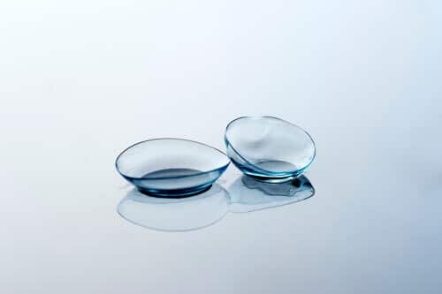
Lead by Dr. John D. Gelles, the contact lens division of the CLEI Center for Keratoconus has unparalleled experience in specialty contact lens fitting for keratoconus and other corneal diseases. Specialty contact lenses are the mainstay of visual improvement for individuals suffering from vision loss associated with keratoconus and other irregular corneal conditions. Our center is exclusively devoted to addressing the needs of keratoconus patients.
Specialty contact lenses are only a part of the management for keratoconus and irregular corneal disease management. Keratoconus is progressive in nature and treatments such as corneal collagen crosslinking are vital to slowing progression. Additionally visual correction procedures for irregular corneas, such as topography guided PRK, may improve uncorrected vision and vision with glasses while improving the cornea symmetry. This shape improvement may make contact lenses simpler.
At the Cornea and Laser Eye Institute and the CLEI Center for Keratoconus. Our doctors are world renowned experts in keratoconus and irregular corneal disease management and research.
They have written many of the foundational manuscripts on keratoconus management. Dr. Hersh was also the principal investigator, medical monitor and primary author on the clinical trials and manuscripts related to corneal collagen crosslinking in the United States. Our doctors collaborate to provide the best care to manage your condition.
The Specialty Contact Lens Design Process at CLEI
The process of designing a customized lens to meet the needs of the keratoconus patient starts with an advanced and in depth evaluation. This evaluation guides the process to understand which type of specialty contact lens will meet your individual needs. A multitude of different scans, impressions and diagnostic lenses may be used in the design process. Once a lens type has been selected, our doctors begin the process of creating a custom contact lens for you. This process generally involves several revisions or modifications to ensure the lens is ideally fit, eye health is maintained and vision is the best possible.
Understanding Vision from Normal Corneas versus Keratoconus and Irregular Corneas
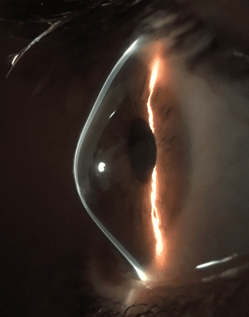
Normal Cornea
The eye is similar to a camera, a series of lenses (cornea and internal lens in the eye) are in place to help light focus on the film or camera sensor (retina in the eye). The cornea is analogous to the front lens on a camera. Normal corneas have a smooth dome shape, this allows light to focus to a single, sharp focal point. In a perfect eye this focal point lands on the retina. If the focal point is in front of or behind the retina the vision is blurry. This misalignment is called refractive error. Glasses, contact lenses, or vision correction surgery can aid in moving the focal point to the retina. This creates clear distortion free vision.
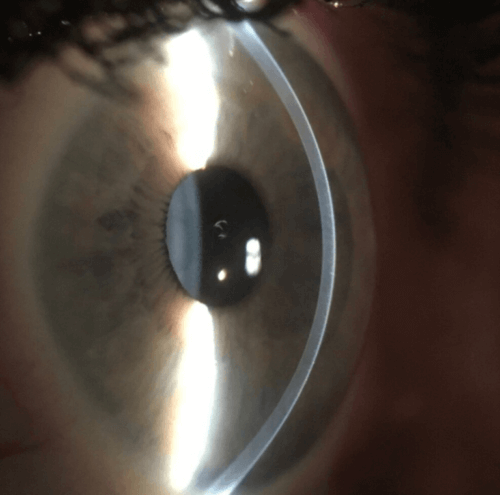
Keratoconus and Irregular Cornea
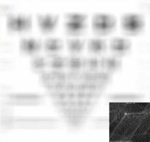
A irregular cornea is one that has lost its smooth dome shape. This can be caused by a variety of conditions such as keratoconus. This irregularity causes light to distort, resulting in multiple focal points. This creates a type of blurred vision that can be described as distorted, overlapping, or ghosted and is also responsible for glare, flare and halos. Glasses and standard soft contact may help in moving the multiple focal points back toward the retina but these focal points remain separate and the vision still distorted. This visual distortion is called an higher order aberration.
Vision Simulations with Glasses Correction
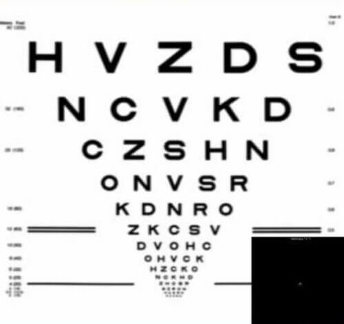
Normal Cornea
Vision Simulation of a healthy eye with a normal cornea wearing glasses. Image by John D. Gelles, O.D. Copyright © 2020 by the Cornea and Laser Eye Institute. All Rights Reserved.
Keratoconus & Irregular Cornea
Vision simulation from an eye with keratoconus wearing glasses. Image by John D. Gelles, O.D. Copyright © 2020 by the Cornea and Laser Eye Institute. All Rights Reserved.
Understanding Vision with a Specialty Contact Lens for Irregular Cornea
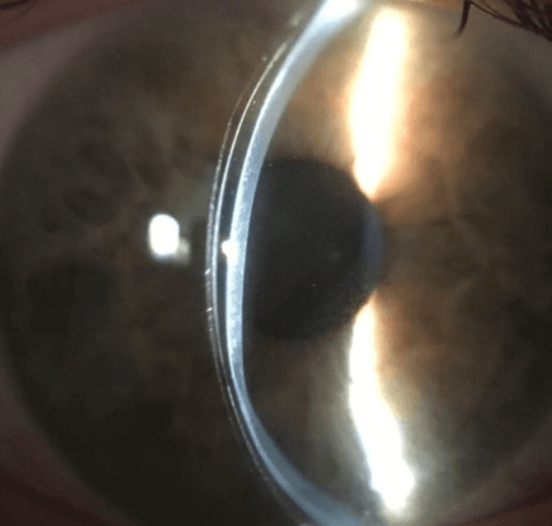
A specialty contact lens masks the irregular cornea shape. The lens surface creates a smooth dome shape, and this allows the multiple focal points to come back into a single focal point, improving vision and reducing distortion.
When a specialty contact lens is worn, the vision is significantly improved. In some cases vision will need to be further refined with the use of highly customized optics, even further improving vision and reducing distortion.
Visual Simulation with a Specialty Contact Lens
Vision simulation of the same keratoconus eye while wearing a scleral lens. Image by John D. Gelles, O.D. Copyright © 2020 by the Cornea and Laser Eye Institute. All Rights Reserved.
Keratoconus and Irregular Corneas
Irregular corneas develop as a result of disease, dystrophy, trauma, or surgical complication. The resultant vision from the asymmetric corneal shape cannot be adequately corrected with spectacles or traditional soft contact lenses. The primary function of specialty contact lenses for this population is to mask the abnormal corneal surface, reducing the associated blur and improving visual acuity.
Irregular Corneal Conditions:
Corneal Ectasia: These are diseases characterized by weak and thin corneal tissue resulting in an irregular cornea shape. Examples include conditions such as keratoconus, keratoglobus, pellucid marginal degeneration, and corneal ectasia after surgery.
Post-Keratoplasty: A keratoplasty is also known as a corneal transplant. These surgeries are used when the cornea is severely scarred or have extremely advanced disease. They can involve the replacement of all or select layers and areas of the cornea. After transplantation the cornea is smoother and clearer but irregularities still exist. Examples include penetrating keratoplasty (PK), deep anterior lamellar keratoplasty (DALK), corneal tectonic grafts (patch grafts), and epikeratophakia.
Post-Corneal Surgery: In very rare cases, after a corneal procedure to reshape the cornea, the corneal surface may become irregular in shape. Examples include after laser assisted in situ keratomileusis (LASIK), photorefractive keratectomy (PRK), radial keratotomy (RK), or intracorneal ring segment (Intacs, Keraring, MyoRing, Ferrara Ring).
Corneal Opacity: A corneal opacity is a scar in the cornea. Scars may result in an irregular corneal surface and can occur after trauma, infection, or advanced ectasia.
Corneal Dystrophies: These are genetic, often progressive, conditions where abnormal tissue often accumulates in the cornea. Anterior and stromal dystrophies such as lattice, granular, Meesmann, Cogan, and Reis-Bucklers dystrophies may result in an irregular corneal shape.
Corneal Degenerations: These conditions cause deterioration and sometimes impair function of the peripheral cornea tissue. Examples include Salzmann’s nodular degeneration and Terrien’s marginal degeneration.

