The cornea is the clear, dome-like outer lens of the eye (like a crystal on a watch). In LASIK, an excimer laser removes tissue from the center of the cornea to flatten its curvature and correct nearsightedness and astigmatism. The laser essentially reshapes the cornea’s front surface. To do this, the corneal stroma (the tissue beneath the corneal epithelium) must be exposed. This usually is accomplished using a second laser, called a femtosecond laser to create what is known as a corneal flap.
The general goal of LASIK is to reshape the cornea so that the rays of light that enter the eye are focused clearly onto the retina. The excimer laser produces a cool, ultraviolet beam of light (193 nanometers in length) that literally vaporizes tissue away as it breaks carbon‑to-carbon bonds without harming adjacent tissue. Tissue is removed in a precise fashion on a microscopic level, leaving adjacent tissue unharmed. This vaporizing process is called photoablation.
The unparalleled precision of the excimer laser makes it uniquely suited to the task of refractive corneal surgery. Each pulse of the laser removes 0.25 microns of tissue. Think of it as slicing 1/200 of a human hair, 1/28 of a red blood cell, or 1/39 millionth of an inch in 4 billionths of a second. This allows the surgeon to literally sculpt the cornea into a more desirable shape, gently and precisely, and allows the rays of light to focus properly on the retina.
In the LASIK procedure, the LASIK flap is first prepared with the femtosecond laser. It is done with local anesthetic drops and a mild sedative, so the procedure is entirely comfortable. The flap is pulled back like a tiny, clear, hinged lid and the corneal stroma is exposed. The laser part of the LASIK procedure takes place in the exposed corneal bed (corneal stroma). The laser application itself (using the Wavelight laser) lasts about thirty seconds. After the exposed corneal bed is treated by the laser, the flap is replaced.
We have been doing LASIK for 25 years. In fact, Dr. Hersh was lead author of the study leading to first FDA approval of laser vision correction in the United States (https://www.vision-institute.com/wp-content/uploads/2018/03/Copy-of-Results-of-Phase-III-PRK-for-Myopia-vol-104-No-10-Oct-1997.pdf). Topography-Guided (TG) LASIK (also known as Contoura LASIK) is the newest advance in laser vision correction. The goal of TG-LASIK is to be even more precise than with previous technologies, to optimize your visual results, and to help avoid night vision side effects such as glare or haloes around lights.
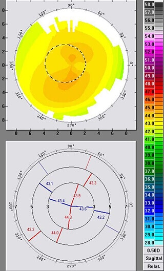
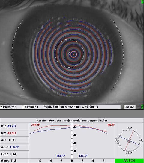
TG-LASIK uses your own customized corneal topography map to plan your treatment. Aside from nearsightedness or astigmatism, vision can also be affected by other individual optical irregularites or aberrations. These aberrations can be thought of as static light, which lessens the clarity of the signal or image. This is analagous to everyday TV use. For instance, a high definition TV has more focused signal and less static or aberrations, and thus a crisper picture. The same is true for your eye. Each eye has a unique aberration profile which remains even if nearsightedness and astigmatism is corrected. After LASIK, specific aberrations or visual static components can potentially affect night vision, with symptoms such as glare and halo images. Minimizing or correcting these aberrations will, we hope, make your vision correction more precise, whether done by a LASIK or LASEK/PRK technique. In addition to your glasses prescription, in TG-LASIK, we also program your individual corneal map into your laser treatment. The corneal topography map is like a map of a mountain range taken from a satellite – red areas are higher (like a mountain), blue areas are lower (like a lake), and green represents a normal slope. By programming you map directly into the laser, our goal is to further remove optical irregularities from your cornea and give you an even better corneal shape with, consequently, improved visual function. TG-LASIK is not for everyone and we will need to evaluate your particular situation to see what’s your best option in vision correction surgery.

